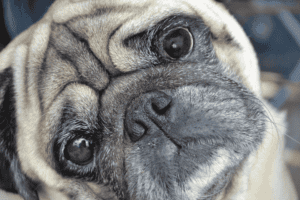
If you’ve recently noticed your pet shaking their head frequently, they may be headed for an ear problem called an aural (ear) hematoma. A hematoma is a collection of blood that is localized in a certain area, in this case, the ear flap of dogs, and less commonly, cats. When a hematoma is present, the pinna (ear flap) will appear thickened, swollen, and spongy, and will be sensitive and warm to touch. Since this is a condition we see often in our urgent care service at our Union location, we thought we’d spend some time discussing what causes aural hematomas, and how we can treat and prevent them.
What causes an aural hematoma?
The ear structure is made up of two thin layers of skin, with a layer of cartilage in between the skin layers. Blood vessels run just beneath the skin. Most commonly, ear hematomas occur when something in your pet’s ear canal irritates them, and they respond with violent head shaking that damages the delicate blood vessels in the ear, causing one or more to break. This results in bleeding into the space between the skin and the cartilage on the inner surface of the ear.
Common causes of irritation of the ear canal include:
- Foreign body (foxtail awn, grass seeds, ticks)
- Ear mites
- Seasonal allergies
- Otitis externa (ear infection)
- Trauma (fighting with other animals)
- Bleeding or clotting disorders (less common)
Treatment
There are several methods for the treatment of aural hematomas, depending on the size and location of the hematoma, your pet’s overall history and general health, and their level of pain or discomfort. Your pet’s veterinarian will discuss treatment options to determine a plan with you, based on these factors.
Some hematomas can reabsorb on their own, and your veterinarian may not recommend treatment if the hematoma is small and does not cause your pet discomfort. However, most larger hematomas are quite uncomfortable for your pet. Allowing the hematoma to reabsorb on its own is a process that may take weeks, and could result in permanent deformity of the ear and possible obstruction of the ear canal. In these cases, medical intervention of some kind is usually recommended.
A corticosteroid injection can be effective at reducing inflammation and encouraging healing, either on its own or in conjunction with other treatments. Cold laser therapy may also be very beneficial to reduce fluid and swelling, speed healing, and manage pain and inflammation.
Your veterinarian may recommend aspiration, which involves inserting a needle into the hematoma and drawing out the blood and fluid using a syringe. Aspiration is a relatively easy procedure to perform and many hematomas resolve with this method. After the procedure, your pet’s ear may be wrapped snugly against their head until healing can occur, sometimes using a protective bandage such as a V-Bonnet or No-Flap Ear Wrap. This prevents the area from re-filling if your pet shakes their head.
Another method is surgical intervention, during which your pet is placed under general anesthesia. One or more surgical incisions are made in the pinna and the hematoma is drained. Sutures are then placed to attach the inner ear cartilage to the outer, eliminating any space for blood and fluid to refill.
Your pet will need to be seen for a medical progress exam and to remove any bandages, drains, or stitches that have been placed. If it is determined that your pet’s hematoma was caused by a foreign body in the ear, an ear infection, allergies, or ear mites, your veterinarian will also monitor this condition and may prescribe medications to treat it.
Aural hematomas can be painful, so if you see your pet shaking their head frequently or notice swelling in the pinna, don’t hesitate to call us right away.